Veröffentlichungen
In-vivo-Studien/präklinische Studien
Bizenjima T, Takeuchi T, Seshima F and Saito A: Effect of poly (lactide-co-glycolide) (PLGA)-coated beta-tricalcium phosphate on the healing of rat calvarial bone defects: a comparative study with pure-phase beta-tricalcium phosphate. Clinical Oral Implants Research (2016). Favero V, Lang N P, Canullo L, Urbizo Velez J, Bengazi F and Botticelli D: Sinus floor elevation outcomes following perforation of the Schneiderian membrane. An experimental study in sheep. Clinical Oral Implants Research (2015).
Schmidlin P R, Nicholls F, Kruse A, Zwahlen R A and Weber F E: Evaluation of moldable, in situ hardening calcium phosphate bone graft substitutes. Clinical Oral Implants Research (2013).
Yip I, Ma L, Mattheos N, Dard M and Lang N P: Defect healing with various bone substitutes. Clinical Oral Implants Research (2014).
Zigdon H, Lewinson D, Bick T and Machtei E E: Vertical Bone Augmentation Using Different Osteoconductive Scaffolds Combined with Barrier Domes in the Rat Calvarium. Clinical Implant Dentistry and Related Research (2012).
Klinische Studien
Dudek D, Sołtykiewicz K, Helewski K, Wyrobiec G, Harabin-Slowinska M, Kowalczyk-Ziomek G and Wojnicz R: Treatment of a mandibular cyst with synthetic bone graft substitute. Implants (2013) 2013(1): 34-36.
El Sayed E, Khalil A and Saleh M: Clinical and radiographical evaluation of immediate implant versus delayed implant after socket preservation of upper anterior teeth. Alexandria Dental Journal (2015) 40: 79-85.
Jurisic M, Manojlovic-Stojanoski M, Andric M, Kokovic V, Danilovic V, Jurisic T and Brkovic B B: Histological and morphometric aspects of Ridge preservation with a moldable, in situ hardening bone graft substitute. Arch. Biol. Sci. (2013) 65(2): 429-437.
Kakar A, Chaudhary V, Kakar R C, Lahori M and Kakar K: Indirect Sinus Elevation And Implant Placement Using A Modified Crestal Approach - A Case Report. The Journal of Academy of Oral Implantology (2011) 3(Jan-Apr): 37-40. Leventis M D, Fairbairn P, Kakar A, Leventis A D, Margaritis V, Lückerath W, Horowitz R A and Nagursky H: Minimally invasive alveolar ridge preservation utilizing an in situ hardening ß-tricalcium phosphate bone substitute. A multicenter case series. International Journal of Dentistry (2016) 2016.
Neumeyer S and Neumeyer-Wühr S: The use of polylactide- coated beta-TCP Closure of oroantral communications. Implants (2010) (4): 32-36.
Thoma K, Pajarola G F, Gratz K W and Schmidlin P R: Bioabsorbable root analogue for closure of oroantral communications after tooth extraction: a prospective case-cohort study. Oral Surg Oral Med Oral Pathol Oral Radiol Endod (2006) 101(5): 558-64.
Troedhan A, Kurrek A, Wainwright M, Schlichting I, Fischak-Treitl B and Ladentrog M: The transcrestal hydrodynamic ultrasonic cavitational sinuslift: Results of a 2-year prospective multicentre study on 404 patients, 446 sinuslift sites and 637 inserted implants. Open Journal of Stomatology (2013) 3: 471.
Troedhan A, Schlichting I, Kurrek A and Wainwright M: Primary implant stability in augmented sinuslift-sites after completed bone regeneration: a randomized controlled clinical study comparing four subantrally inserted biomaterials. Scientific reports (2014) 4.
Troedhan A, Wainwright M, Kurrek A and Schlichting I: Biomechanical Stability of Dental Implants in Augmented Maxillary Sites: Results of a Randomized Clinical Study with Four Different Biomaterials and PRF and a Biological View on Guided Bone Regeneration. BioMed Research International (2015) 2015
Klinische Fälle
Kammerhaltung
| Indikation | Kammerhaltung |
| Patient | 23 Jahre alt, weiblich |
| Position | Zweiter Prämolar im rechten Oberkiefer (15) |
| Verwendetes Material | GUIDOR® easy-graft CLASSIC |
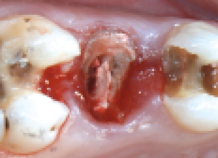
Zweiter Prämolar im rechten Oberkiefer (Zahn 15) mit Karies.
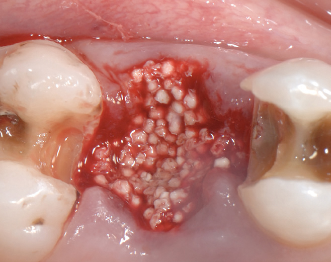
Kammerhaltung mit easy-graft CLASSIC nach atraumatischer Extraktion.
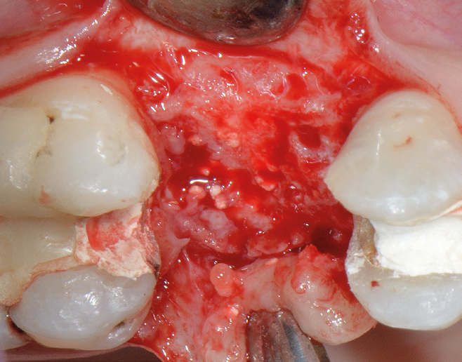
Situation bei der Wiedervorstellung 4 Monate nach der Operation, easy-graft CLASSIC-Granulat ist gut in den neuen Kochen integriert.
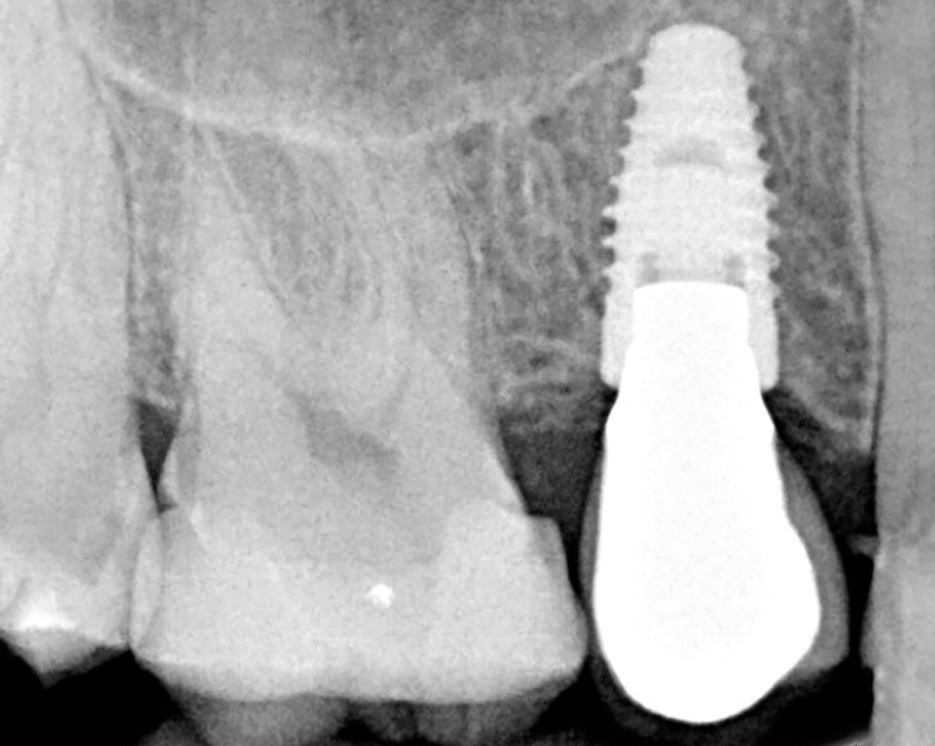
Letzte Röntgenaufnahme 16 Monate nach der Operation.
Dr. Minas Leventis
Periimplantäre Knochengeneration, unmittelbare Implantation
| Indikation | Periimplantäre Knochengeneration, unmittelbare Implantation |
| Patient | 45 Jahre alt, weiblich |
| Position | Mittlerer Schneidezahn im rechten Oberkiefer (II) |
| Verwendetes Material | GUIODOR® easy-graft CRYSTAL |
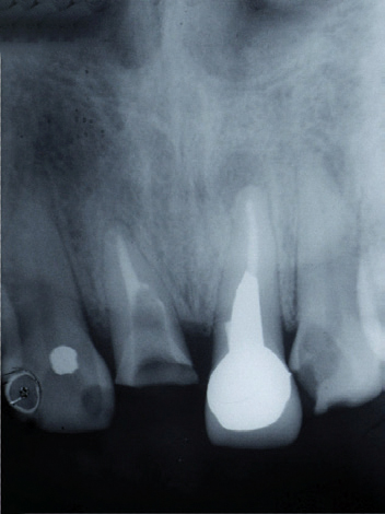
Röntgenbild der Ausgangssituation.
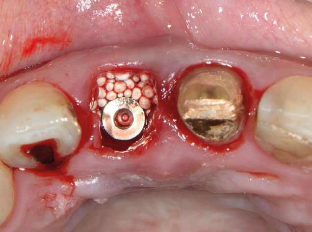
Unmittelbare Implantatinsertion und Transplantat.
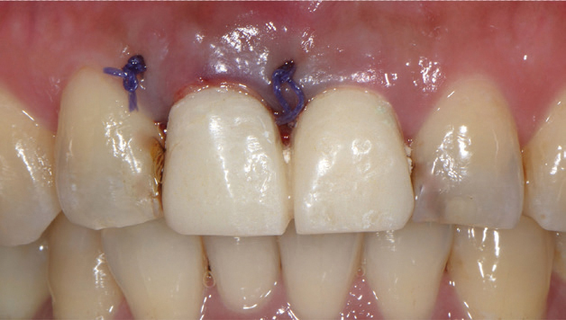
Unmittelbare provisorische Versorgung.
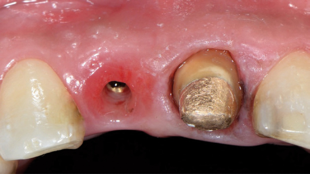
5 Monate nach der Operation, ausgezeichnete Erhaltung der Kammarchitektur.
Dr. Minas Leventis
Laterale Sinusboden-Augmentation
| Indikation | Laterale Sinusboden-Augmentation |
| Patient | Fallserie von 20 Sinusboden-Augmentationen |
| Position | Oberkiefermolare |
| Verwendetes Material | GUIDOR® easy-graft CRYSTAL |
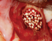
Laterale Sinusboden-Augmentation mit easy-graft CRYSTAL.
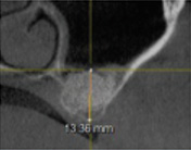
Kontroll-CBCT nach 6 Monaten.
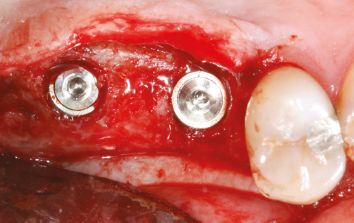
Implantation nach 6 Monaten. (Position 16, 17).
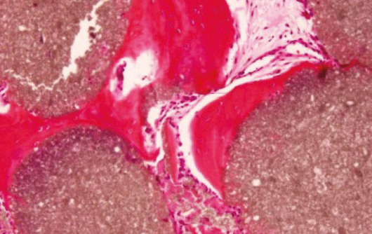
Histologische Aufnahme zeigt in neu gebildetem Knochen eingebetteten easy-graft CRYSTAL.
Dr. Antonio Flichy-Fernández